Regardless of what you would possibly believe, the most effective neurologist in Panchkula and neurology hospitals aren’t superheroes. They don’t have X-ray eyesight, nor will they read heads and determine what’s happening under the skin without appropriate assessment. Assessment includes that which you inform them about your injury or illness and therefore the evaluation of bodily movements that excite your own symptoms. By doing this, they create theories of possible causes impacting your injury or illness. Sometimes, they’d send you to induce a scan to gauge your internal arrangements to affirm or deny our notions.
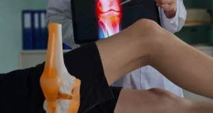
Scans, otherwise called medical brain imaging aids in looking under the surface and distinguishing influenced structures inside the body causing the injury. they’re rather cheap and accessible due to the best neurologist in Panchkula that gives imaging services together with the advancement in medical engineering.
The easy answer is that the bulk of patients don’t need to become imaging thanks to the character of the injury/condition. Just a minority of people need extra medical imaging to assist within the diagnosis/management procedure. This relies upon a range of matters that the individual has stated about the background of this illness and what the most effective neurologist in Panchkula shows us with motion.
WHAT’S BRAIN IMAGING?
Brain imaging also observed as neuroimaging, is the procedure of analyzing the mind and also the system with various imaging methods. There are several different brain imaging scans that provide either an on the spot or an indirect picture of your brain.
Structural imaging means brain scans which analyze the development of the system. It’s often utilized to diagnose intracranial disorder and harm to a number of their brain’s structures.
WHO MUST PROCEED FOR BRAIN IMAGING?
Brain imaging is advantageous for people suspected of getting difficulties or medical conditions impacting their own brains. On the opposite hand, the identification and therapy of neurological disorders is barely one intention of brain imaging. The method also helps the most effective neurologist in Panchkula to understand the aim of the assorted areas of the mind and also conduct research and build new technology for treating brain disorders. The brain disease types are:
- Structural diseases
- Tumors
- Bleeding
- Blood clots
- Inflammation
- Birth defects
- Stroke
- Functional ailments
- Alzheimer’s disease
- Epilepsy
- Dementia
- Memory ailments
Brain ailments can cause several symptoms or complications, such as:
- Vision reduction
- Weakness
- Paralysis
- Compromised brain functioning
Before sending a patient to medical imaging, the best neurologist in Panchkula should first understand whether the patient fits the standards for medical imaging. this can be done by weighing variety of great variables:
IMAGING ALGORITHM
Does this harm tick the boxes suggested for this collection of images to be achieved? for example within the case of getting a sprained ankle. Before an X-Ray may well be taken the individual should clarify pain on the bony edges of your ankle or not be able to square on the ankle either right after the injury occurred or once they pose for a clinical examination. If the patient doesn’t meet the suggested standards it’s not going that the image requested are of any advantage to the identification procedure. The plan also directs another course of therapy when the image comes back negative or positive for an ankle break. The most effective neurologist in Panchkula would normally describe this procedure for you before sending you away to urge an image.

SORT OF IMAGE
Based on the type of harm another type of picture could possibly be critical. The styles of cells they’re inquisitive about detecting are bone, bone, ligament, cartilage, tendons and nerve-wracking. Every type of picture is particular to specific cells in contrast to a distinct quiet medical imaging.
X-Ray: the image is created by exposing the goal region to an electromagnetic wave called X-Rays. The X-Rays have the body and render a shadow of rectal tissue since it’s impossible for them to labor under the dense arrangement of bone. X-Ray is useful for diagnosing dislocation or fracture of bones, they’re usually utilized as a primary measure to rule out severe harm which may involve surgical intervention.
Ultrasound: Ultrasound is generated by introducing a solid wave into the structures immediately under the epidermis. This picture aids in identifying fractures in normal cells and or swelling within the affected region. This works great for delicate tissues like tendons and ligaments but isn’t quite as precise as other forms of images. Ultrasound is usually appropriate since it’s a more cost-effective alternative to MRI.
CT (Computed Tomography): The device performs multiple X-Rays round the affected area in a very circular way thanks to making a 3D image on a pc. The images are displayed as cross-sectional”slices” that reveal various tissues in various reminder gray. CT’s are helpful at demonstrating minor cracks in bone tissue and changes in tissue balancing identifying harm to the tissue. CT is often utilized as a substitute for MRI because there’s not any magnetic flux made by the machine and its functionality.
MRI (Magnetic Resonance Imaging): Patients are expected to place still as they’re introduced into a narrow tube comprising the scanning equipment that emphasizes fat and water molecules within the body. These compounds in various proportions represent different tissues within the body. Damage and injury can be seen as disruptions into the results of the tissues and notify clinicians of abnormality inside the tissue.
RADIATION
Particular forms of medical imaging are conditional on the usage of radiation to form pictures. X-rays and CT demand the usage of X-Rays that sit at the highest end of the spectrum. With increased energy that this sort of radiation has the facility to influence normal atom behavior within the entire body, this can be called radiation. Changes in Biology behavior has a control on the electron cloud of the molecule, altering the quantity of electrons connected to the atom. This might result in cell mutation that has the aptitude to spice up the probability of cancerous cell growth within the body.
POTENTIAL RISKS AND COMPLICATIONS
The various types of brain imaging scans include their own set of dangers and pitfalls. That’s the rationale why the bulk of neuroimaging scans are utilized with caution by the most effective neurologist in Panchkula. On the flip side, an MRI scan isn’t safe to be used on patients with metallic devices implanted in their bodies, sort of a pacemaker.
MRI scans also need patients to lie still. Hence, this can be not an honest idea for stubborn patients. The strategy by which the method is completed also carries some dangers for patients that are claustrophobic.
For the other information regarding neuroimaging, you’ll contact JP Hospital Zirakpur, the best neurosurgery hospital in Chandigarh. This hospital has the best neurologist in Panchkula, which may facilitate your in solving every neuro-related issue.